Thursday, January 30, 2014

Have you twisted your ankle… again?

We’ve all been there. That horrible moment when you miss a step and fall awkwardly off the curb, or are tackled from the side with your foot planted (usually on the sports field, but not necessarily), feeling that awful snap in your ankle. High heeled shoes and icy weather have a lot to answer for too. Now we can’t go attributing twisted ankles to wearing high heels all of the time, but certain footwear – and certain activities – do seem to cause more problem s than others


  • The most common injury reported with a “twisted ankle” is a sprain to the lateral ligaments of the ankle (the ones on the outside as opposed to the inside [medial]). Normally the symptoms will include swelling, pain, bruising, heat and redness, along with stiffness over a period of time.
  • One of the big differences between ligaments (which hold bones together) and muscles is that muscles are contractile, meaning that you can actively and purposefully shorten them. By over-working a muscle, you may strain it (note that’s “strain” with a “t”). Ligaments are generally stiffer (thankfully, otherwise our bones would be wobbling all over the place), but when we stretch them too far, we can sprain them (with a “p”).
  • Let’s start with a diagram of the bones of the foot. The ones we’ll be concentrating on are the tibia and fibula (which make up the lower leg), the talus and the calcaneus (heel). The other bones in the foot are important for movement, but not strictly involved at the ankle joint.

44


  • The ankle joint itself is formed by the tibia (the shin bone), the fibula (the smaller lower leg bone) and the talus. It  is a hinge joint, meaning it moves up and down (dorsiflexion and plantarflexion) but not side to side – these movements are prevented by the strong medial and lateral ligaments, the medial being stronger than the lateral (which is one reason we tend to injure the lateral ones more often).
The lateral ligament is actually split into 3 parts (see the diagram below): the anterior and posterior talofibular ligaments and the calcaneo fibular ligament. The most commonly injured part is the anterior talofibular ligament.


There are 3 types of ligament sprain, each varying in intensity.

Grade I sprains are those with very little damage. You’ll see some local swelling and feel a moderate amount of pain, particularly if you twist whilst standing on the injured ankle. However, this should ease off fairly quickly following the advice below.


A grade II sprain involves damage to more of the fibers of the ligaments, therefore the swelling will be more pronounced and will appear much more quickly than in a grade I. You’ll be in more pain (sorry about that), and may find it difficult to weight-bear on the injured foot. The good news is that the joint itself remains relatively stable, and the ligaments are still able to hold the bones in place.

The most severe sprain is a grade III. This is a complete rupture of the ligament, meaning that the bones at the ankle joint will lose some of their stability. There will be a lot of swelling and, unfortunately, a fair amount of pain too.

The best way to determine how severe your ankle sprain is, is to have your physiotherapist assess the injury.

  •  So what do you do with a twisted ankle?

The main aims of treatment will be to reduce the pain and swelling in your ankle and to help the tissue healing begin. There are lots of things you can do to facilitate this by using the PRICE mnemonic.

  • P is for Protect. This means be aware of your injury and protect your ankle by further damage e.g. by using crutches, or by keeping the weight off of the injured side as much as possible. (In other words, come off of the pitch and wear sensible shoes for the time being.)
  • R is for Rest. When an injury is sustained to a ligament, some of the small blood vessels supplying the tissue will break. This starves small parts of the tissue of oxygen and nutrients, meaning that the tissue necrosis or dies. It is important to rest as much as possible to limit the amount of oxygen required and therefore limit how much of the tissue dies. Larger injuries may require total rest as your body may be in shock. Smaller injuries will require rest of the affected ankle, but otherwise allow you to continue activity. As your healing continues, the periods of rest will be reduced.
  • I is for Ice. Making the affected area cold will reduce the swelling in the area and lower the metabolism of the tissues – in short this means the tissues need less oxygen, and so the rate of tissue death slows. This is all good for recovery. If you’ve had an injury, the best thing to do is use ice or an ice pack, wrap it in a moist tea towel or use a barrier between the ice and skin (very important to avoid ice burns) and place that over the painful area for 15 minutes every 2 hours or so throughout the first 2 days.
  • C is for Compression. You’ll be well aware by now that ankle injuries tend to swell. By using a simple elastic bandage such as tubigrip [or] crepe bandage, you can provide a uniform compression around the foot, ankle and lower leg to reduce the accumulation and spread of the swelling, which is very common in this type of injury partly due to gravity allowing fluid to pool at the foot.
  • E is for Elevation. you were waiting for this. I’m not one to encourage you to put your feet up… except in this case. Normally the shifting of fluid from the legs is aided by the pumping of the calf muscles, but when you are restricted to rest, the excess fluid from the injury tends to gather around the ankle. Using a stool to place your foot on, this will encourage rest and protection, as well as help drain the excess fluid back up the leg and towards the heart


                               Wrapping crepe bandage


Grade I sprains can be treated solely with the PRICE first aid, however grades II and III strains will likely need some form of support such as taping, or a cast or splint, and some solid advice on how to deal with the injury, particularly in the early stages of recovery

  • Use of electrotherapy such as ultrasound and laser treatment can reduce pain and inflammation and promote healing

ankle taping procedure. The superior anchor(second photo)   was applied in a standardized way according to the subject's body dimensions,at 35% of the distance from the lateral malleolus to the fibula head.


  • Once the swelling has started to ease slightly (generally after the first day or 2 for grade I sprains) you can begin some gentle exercises to help your ankle regain normal movement. Your physiotherapist can help you by designing a specific programme for you which will help you recover from injury and return to your normal activities as soon as possible, as well as helping you avoid future ankle sprains (even if you really MUST wear those high heel


 Disclamier;This information might have been copied from different sources to give the best accessible


Wednesday, January 29, 2014

Basic Stretching Program

Stretching 

Stretching or lengthening your muscles helps you become more limber, which makes certain activities of daily living easier. Plus, the improved flexibility gained from stretching regularly helps you prevent injuries when you are performing everyday body movements and especially during exercise. Stretching also reduces muscle tension, increases circulation, improves posture, and it just naturally feels good! 

The FITT principle can help you incorporate stretching exercise into your physical activity plan.

  • Frequency (how often you are physically active in a week)

Aim to do stretching exercises as many times as you do cardio—working toward five times per week.  Stretch all the major muscle groups. The more frequently you stretch, the more quickly you will improve your flexibility.

The best time to stretch is usually after you cool down at the end of a cardio exercise session. At the very least, warm up by walking for 5 minutes before stretching. Stretching after you’ve increased blood flow to your muscles and after your tendons and ligaments have been in use will minimize possible injury during stretching.

  • Intensity (how hard you work each time you are physically active)

Each stretch should be performed with a slow, steady movement without bouncing or locking your joints, which can cause injury. Stretch just to the point of mild discomfort, stopping before the stretch becomes painful.

  • Time (the duration or how long your physical activity lasts)

Aim to stretch for 10 to 15 minutes at a time. Hold each stretch for about 15 seconds. Repeat as necessary, according to what feels good.

  • Type (the kind of physical activity you are doing)

There are many different stretches you can do to help increase the range of motion of all your joints.

Common Stretches


Hamstring Stretch

                                   Quadriceps Stretch [Standing Quad Stretch]

Quad Stretch

                        Inner Thigh and Hip Stretch [Seated Butterfly Stretch]

Inner Thigh and Hip - Seated Butterfly

                                  Abdominals Stretch [Lying Abs Stretch]

ABdominals Stretch

                          Lower Back Stretch [Lying Double Knee Stretch]

Lower Back Stretch

                                Upper Back Stretch [Cat Stretch]

Upper Back Stretch

                              Chest Stretch [Standing Chest Stretch]

Chest Stretch

                            Shoulders Stretch [Standing Shoulder Stretch]

Shoulders Stretch

                                 Triceps Stretch [Standing Triceps Stretch]

Triceps Stretch

                         Biceps Stretch [Standing Wrist-Biceps Stretch]

Biceps Stretch

                                                      Neck Stretch

Neck Stretch


Interval Training

Interval training involves simply alternating a low-intensity activity (such as walking) with short bursts (for example, 30 to 90 seconds) of a higher-intensity activity (such as running) throughout your workout session.

The Benefits of Interval Training:

  • Interval training boosts the calories you burn. So, you can burn more calories in less time, which we all are short on!

  • Interval training improves your cardiovascular endurance, or aerobic capacity. This helps your heart and lungs function more efficiently.

  • Interval training keeps your body challenged.

  • Interval training helps pass the time you are exercising by keeping your mind busy during the activity.

How to Begin Interval Training:

  • First check with your doctor, because interval training is not for everyone. If you have a chronic health condition or have not been exercising regularly, you may not be able to tolerate interval training.

  • Once you have clearance from your doctor, it is a good idea to begin interval training slowly.Try adding only one or two bursts of a higher–intensity activity in your exercise session.  Then increase the number of bursts in future sessions as tolerated.

    • If you’re a novice, try walking and then adding 30 to 60 seconds of walking faster. Then return to a slower walking pace. Repeat if tolerated.

    • If you’re in good shape, try walking more briskly and then adding 30 to 90 seconds of light jogging, as tolerated. Then return to a brisk walk. Repeat if tolerated.

  • Get guidance. Consider meeting with a physical therapist, exercise physiologist or certified personal trainer, who can recommend and oversea an interval training schedule so you get the most out of your workouts

  • Disclaimer:This information might have been copied from different sources to give the best accessible

Tuesday, January 28, 2014

Osteo arthritis of knee/osteo arthrosis/Degenerative joint disease(kneepain)


While age is a major risk factor for osteoarthritis of the knee, young people can get it too. For some individuals, it may be hereditary. For others, osteoarthritis of the knee can result from injury or infection or even from being overweight.
Osteoarthritis, commonly known as wear and tear arthritis, is a condition in which the natural cushioning between joints -- cartilage -- wears away. When this happens, the bones of the joints rub more closely against one another with less of the shock-absorbing benefits of cartilage. The rubbing results in pain, swelling, stiffness, decreased ability to move and, sometimes, the formation of bone spurs.  


Pathogenesis of Osteoarthritis


Signs:


  • Pain on movement

  • Swelling of the joint

  • Articular gelling - stiffness lasting short periods and dissipates after initial ROM.

  • Spasm of periarticular muscles

  • Bony enlargement of the joint

  • Limitation of range of movement - End ROM mostly affected

  • Crepitus when moved - loud cracks & crunches if the arthritis is severe.

  • Tenderness to pressure

    • Around the joint margins

    • Very sore if knocked or injured in any way

  • Joint Deformity or poor alignment

    • Genu Varum (Bow Legs)

    • Genu Valgum (Knock Knee)
Symptoms: 

Pain:

The types of pain are aching: fleeting and constant, refered: passing down the limb, Sharp stabbing: loose bodies, throbbing: mainly at night. Or bursitis.
    • Early Disease Condition - Gradual onset, mild in intensity, brought on by joint usage, and relieved with rest.

    • May be vague around joint

    • Self-limited or intermittent

    • Severe Disease Condition - pain at rest & during night

    • Severity of pain does not reflect the severity of the disease

  • Joint stiffness < 30 minutes in the morning and becomes worse as the day goes on.

  • Loss of normal function of the joint and the person as a whole.

  • Advanced Condition - Sense of grinding or locking of a joint, and buckling or instability of joints during demanding tasks.
Modifiable risk factors

Obesity, Occupation, Trauma, Sports


Non-modifiable risk factors


Age; gender (female); ethnicity; family history.


CONSERVATIVE TREATMENTS & PHYSICAL THERAPY ADVICE


The primary goals of treating osteoarthritis of the knee are to relieve the pain and return mobility. The treatment plan will typically include a combination of the following:


Pharmological therapy:
  • Pain relievers and anti-inflammatory drugs. This includes over-the-counter choices such as acetaminophen (Tylenol), ibuprofen (Advil, Motrin), or naproxen sodium (Aleve). Don't take over-the-counter medications for more than 10 days without checking with your doctor.

  • Taking them for longer increases the chance of side effects. If over-the-counter medications don't provide relief, your doctor may give you a prescription anti-inflammatory drug or other medication to help ease the pain.

  • Glucosamine and Chondroitin Sulfate may prove beneficial in alleviating the symptoms    and complaints of symptomatic osteoarthritis of the knee.

  • . Chronic use of glucosamine chondroitin sulfate may elevate serum cholesterol and we encourage you to discuss its use with your Primary Care Provider.

Injections of corticosteroids or hyaluronic acid into the knee

Steroids are powerful anti-inflammatory drugs. Hyaluronic acid is normally present in joints as a type of lubricating fluid. Alternative therapies. Some alternative therapies that may be effective include topical creams with capsaicin


Non-pharmological therapy:
  • Using devices such as braces, There are two types of braces: "unloader" braces, which take the weight away from the side of the knee affected by arthritis; and "support" braces, which provide support for the entire knee.

  • Occupational therapy Occupational therapists teach you ways to perform regular, daily activities, such as housework, with less pain.
Physical therapy:
  • Physiotherapy is a non-pharmacological conservative treatment approach that is recommended in clinical guidelines for the management of knee osteoarthritis.

  • Physiotherapy should be considered prior to pharmacological interventions for patients with osteoarthritis, given its low risk of adverse effects and the widespread benefits it can offer most patients with osteoarthritis.

  • Physiotherapy treatment of knee osteoarthritis may encompass any or all of the following: advice, information & education, exercise programs, joint mobilization, muscle re-education, assistance with use of aids [eg walking sticks, braces, orthotics) or electrotherapy modalities. For optimal benefits, physiotherapy management should be based on an individual clinical assessment and tailored to the patient's individual presentation and problems
AIMS:
  1.  To control pain.

  2.  To decrease the swelling.

  3.  To increase the blood supply.

  4.  To prevent further strain or damage to affected joint.

  5.   To improve movement.

  6.   To correct the deformity and contracture.

  7.   To improve muscle power.

  8.  To maintain or improve functional independence

EXERCISE THERAPY


1)Exercise program should be simple.

2)It should be developed on the basis of the normal  kinematics to regain the correct mechanism of knee joint.

3)Exercise program includes.


 Isometric exercises:
  • This is exercise in which muscles are tensed for a period without actually moving them. It can be performed without actually bending a painful joint. As muscles are exercised against resistance, their size and power will increase
QUADS ISOMETRIC EXERCISES
SHORT ARC EXTENSIONS
  • Lie or sit with a rolled towel underneath the knees. Relax the head.

  • Straighten the knees, bringing the heels up off the floor. Keep them up for a count to five

  • Relax. Repeat routine 10 times


SIMPLE ACTIVE KNEE EXTENSION EXERCISE


1 Quad sets

Place a pillow lengthwise under your knee. Pull your toes back and push your leg down to squash the pillow. Hold for 10 seconds and repeat up to 20 times. You should feel your thigh and gluteus muscles during this exercise. Begin with moderate effort and then increase your effort gradually. This exercise can be done lying flat as shown, sitting in a recliner, or leaning against a wall.


2 Hamstring sets

Lie on your back with knees bent and toes up. Dig your heels into the ground and pull back without sliding your heels. Hold for 10 seconds and repeat up to 20 times. You should feel the back of your thigh, the hamstrings, during this exercise. As with quad sets, start with a moderate effort and increase your effort gradually.


3 Straight leg raise

Tighten your thigh and pull your toes back just as you do for the quad set, then maintain the muscle tightness as you slowly raise your leg 18 inches. Lower slowly, relax your muscles briefly, then tighten your thigh, pull your toes back and repeat 20 times.Note: These first 3 knee exercises are the least stressful on the knee joint as they require little to no movement of the knee itself.


4 Bridging

Knees bent and feet flat on the floor as above with your heels spaced comfortably from your bottom. Fold a bed pillow in half, and place it between your knees. Squeeze the pillow and hold on to it tightly. Raise your bottom upward slowly lifting one vertebra at a time from your exercise mat. Continue lifting your bottom upward until there is a straight line from your shoulders to your knees. Hold this position 5-10 seconds. Repeat 6-8 times. Think of reaching your knees forward over your ankles.

Note: If this exercise is uncomfortable, just squeeze the pillow and hold it 10 seconds. This is called an adductor set, as it works the inner thigh muscles. Once you can do this (and knee exercises #1 and 2) 20 times with a strong effort, try bridging again.


5 Knee extension

Sit upright in a chair, straighten your leg, tighten your thigh and pull your toes back. Hold for 10 seconds and repeat up to 20 times. The more you tighten your muscles, the better results you’ll get from this exercise. You may feel a stretch behind your knee during the exercise. For a greater challenge, straighten both legs at the same time, or do this exercise with an adjustable ankle weight up to 5 pounds.

If you feel a strong stretch behind your knee during the exercise, pump your ankle (point your toes and flex your foot back) several times instead of just holding your toes back--this will help improve your flexibility. 


Note: Knee exercises #6 and 7 are balance exercises.

6 Heel raises

Hold onto a chair or countertop only as much as you need to for safety. Go up on your toes as high as possible without leaning your body weight forward. Stay up and maintain your balance for 2-3 seconds. Repeat up to 25 times. Keep your weight over your big toes as you go up and down .Once you can easily do 25 repetitions, try lowering back down on one foot. Then progress to single leg heel raises.


7 Butt kicks

Keep your knees in line and lift your heel as high as possible. Stand upright when you lift your heel. You will feel your hamstrings during this exercise, and you may feel a stretch in the front of your thigh. Repeat up to 20 times on each side. Add an ankle weight to increase the challenge.


   GAIT RE-EDUCATION:


The gait should be analyzed and corrected as required. Gait re-education includes management of stairs, slopes, standing to sitting and bed to chair transfers. Postural awareness needs to be stimulated and the patient encouraged to assume a more efficient posture.

SELF MANAGEMENT:


This includes:

1)Weight control

2)Stress control.

3)Joint protection.

4)Assistive devices.

WEIGHT CONTROL:

  • Weight control is important to successful osteoarthritis management. Being over weight is a risk factor for osteoarthritis. Controlling weight can lessen pain by reducing stress on individual. Weight loss should be occupied with regimen of more Physical Activity. 30 minutes of daily exercise is enough.

    • Stress control techniques are helpful to regain a sense of control while relieving their osteo arthritic pain.

    • Techniques of stress management.

    • Muscle relaxation.

    • Controlled breathing.

    • Biofeedback.

    • Self hypnosis.

    • Time management.
STRESS CONTROL:
 JOINT PROTECTION

  • Joint protection begins with learning new ways to use the    osteoarthirtis of knee joint.

  • Joint stress and strain on the knee joint is limited by following ways.

  • Bend at the knees and straighten the legs.

  • Get up from a chair by sliding forward to the chair’s edge, keeping the feet flat on the floor, and using the palms of the hands to push against the chair’s arms or seat. Stand up by straightening the hips and knee. Use higher seats rather than deep, soft sofas.

  • Never squat or kneel, as these positions strain the hips and  knees.

  • Maintain good posture to avoid putting stress on the joints.

  • Wear well-cushioned athletic shoes with good arch support whenever possible. If dress shoes must be worn, women should choose styles with heels that are no higher than one inch.
ASSISTIVE DEVICES
  • Walking aids like canes, sticks are used.

  • If one knee is effected a single stick is used in the opposite hand.

  • If both the knees are effected tow sticks will be needed.

  •  In severe cases orthotics may be needed.

Surgery: When other treatments don't work, surgery is a good option.

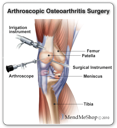
  • If your doctor wants to treat the osteoarthritis in the knee with surgery, the options are arthroscopy, osteotomy, and arthroplasty.

Arthroscopy uses a small telescope (arthroscope) and other small instruments. The surgery is performed through small incisions. The surgeon uses the arthroscope to see into the joint space. Once there, the surgeon can remove damaged cartilage or loose particles, clean the bone surface, and repair other types of tissue if those damages are discovered. The procedure is often used on younger patients (55 years old and younger) in order to delay more serious surgery.


An osteotomy is a procedure that aims to make the knee alignment better by changing the shape of the bones. This type of surgery may be recommended if you have damage primarily in one area of the knee. It might also be recommended if you have broken your knee and it has not healed well. An osteotomy is not permanent, and further surgery may be necessary later on.


  • Joint replacement surgery, or arthroplasty, is a surgical procedure in which joints are replaced with artificial parts made from metals or plastic. The replacement could involve one side of the knee or the entire knee. Joint replacement surgery is usually reserved for people over age 50 with severe osteoarthritis.

  •  The surgery may need to be repeated later if the joint wears out again after several years, but with today's modern advancements most new joints will last over 20 years. The surgery has risks, but the results are generally very good.


Disclaimer: This information might have been copied from different sources to give the best accessible